Eustachian Tube Dysfunction and Neck Pain: Why Your Ears and Your Neck Are Connected
By Dr. Rusty Lavender, D.C. — upper cervical chiropractor, Lavender Family Chiropractic, Sarasota, FL. Reviewed and updated October 2026.
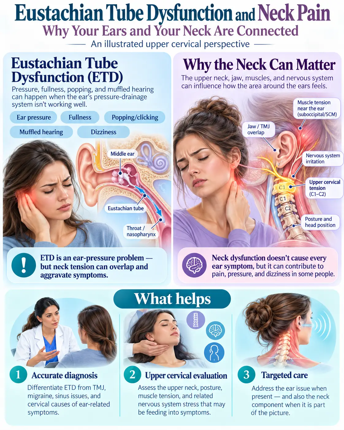
The ear is full. The neck is tight. And nobody has connected the two. If you have Eustachian tube dysfunction — the plugged, underwater, won’t-pop ear that comes and goes for months — there’s a good chance you also have a stiff, aching neck, pressure at the base of your skull, or a headache that wraps forward from behind the ear. You’ve probably mentioned both to the ENT, and the ENT, reasonably, treated the ear. You’ve probably mentioned both to your primary doctor, who treated the neck as a separate problem. They’re not separate. They share nerve wiring, they share a muscle, and they share the same few inches of anatomy at the top of the spine.
This article explains the connection: how the Eustachian tube actually opens and which nerve controls it; why that nerve and the upper neck feed the same brainstem relay; what the research says about ear symptoms referred from the neck; how to tell whether your ear fullness is coming from the tube, the neck, or both; and what an upper cervical evaluation adds to an ETD workup that has already found the ear itself intact.
This is educational content, not medical advice. Ear fullness with sudden hearing loss, severe pain, discharge, fever, or facial weakness needs prompt medical care. Neck pain with fever, stiff neck, weakness, or numbness is urgent.
What Eustachian Tube Dysfunction Is
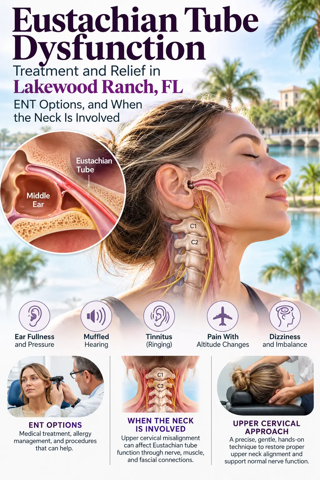
The Eustachian tube is the narrow channel that connects the middle ear to the back of the throat. It’s closed at rest and opens briefly when you swallow, yawn, or chew, letting air in to equalize the pressure behind the eardrum. When it doesn’t open properly, pressure builds, the eardrum is pulled inward, and you get the familiar symptoms: fullness, muffled hearing, popping and crackling, pressure that won’t clear, sometimes mild imbalance.
The international consensus statement by Schilder and colleagues (2015) defined Eustachian tube dysfunction and sorted it into types — obstructive, where the tube doesn’t open enough; patulous, where it stays open too much; and baro-challenge-induced, where it fails under pressure changes like flying or diving. Our Eustachian tube dysfunction guidecovers the types, the symptoms, and the full picture.
The part most ETD explanations skip is how the tube opens. It doesn’t open on its own. A muscle opens it, and that muscle has a nerve, and that nerve has a story.
The Muscle That Opens the Tube — and Its Nerve
The tube’s active opener is a small muscle called the tensor veli palatini. It runs from the base of the skull alongside the cartilaginous part of the Eustachian tube down into the soft palate. When you swallow, it contracts, and its pull on the tube wall is what pops the tube open for a fraction of a second. Leuwer (2016), reviewing the anatomy of the Eustachian tube, describes the tensor veli palatini as the principal dilator, and Alper and colleagues (2012) measured its electrical activity directly and showed that the tube’s opening tracks the muscle’s contraction.
Here’s the key: the tensor veli palatini is controlled by the trigeminal nerve — specifically its mandibular branch, the same nerve that runs your chewing muscles and carries sensation from your jaw, teeth, and the side of your head. The tube’s dilator isn’t on the ear’s nerve supply. It’s on the trigeminal system.
That matters because of what the trigeminal system is connected to.
Shared Wiring: The Trigeminocervical Complex
The trigeminal nerve doesn’t terminate in isolation. Its sensory fibers descend into the brainstem and converge in a structure called the trigeminocervical complex, where they share relay cells with the sensory fibers from the upper cervical nerve roots — C1, C2, and C3, the nerves of the top of the neck. Bartsch and Goadsby (2003) laid out this convergence in their review of the trigeminocervical complex: the brainstem cannot fully distinguish input from the face and head from input from the upper neck, because they arrive at the same cells. It’s the reason an upper-neck problem can be felt as a headache behind the eye, and the reason neck pain is one of the most common features of migraine.
Now connect the pieces. The muscle that opens your Eustachian tube runs on trigeminal signal. The trigeminal system shares its brainstem relay with the upper cervical nerves. When the upper neck is irritated — a misaligned atlas, a whiplash injury, chronic forward-head posture loading the top of the spine — it feeds abnormal input into that shared relay. The trigeminal output that governs the tensor veli palatini is affected downstream. The tube that should open on every swallow opens poorly, or late, or unevenly, and the ear fills.
That’s the mechanism. The neck doesn’t block the tube. The neck disturbs the signal to the muscle that opens it.
Ear Symptoms Referred From the Neck
There’s a second, more direct connection, and the radiology literature documents it well. The ear receives sensory nerve fibers from several sources, including branches of the upper cervical nerves — which is why problems in the neck can be felt in the ear even when the ear is fine. Norris and Chen (2020), in the American Journal of Neuroradiology, mapped the referred-pain pathways to the ear and listed the cervical spine among the sources of secondary ear pain. Ramazani and colleagues (2023), writing for family physicians, note that a large share of ear pain seen in primary care has no ear cause at all — it’s referred from the jaw, the throat, or the neck.
So a person with an irritated upper neck can have two things at once: an ear that genuinely isn’t equalizing well, because the tube’s dilator signal is disturbed, and a sense of fullness or ache in the ear that is itself referred from the neck. Both come from the same place. Both get missed when the ear is examined on its own.
The Clues That Point to the Neck
Not every ETD case involves the neck. Allergies, colds, sinus inflammation, and reflux cause plenty of obstructive ETD with no cervical component. The clues that the neck is part of your picture:
- Neck pain, stiffness, or tightness that travels with the ear fullness — worse together, better together.
- Pressure or pain at the base of the skull, on the same side as the worse ear. Our article on pain and pressure at the base of the skull covers that location.
- A one-sided, stubborn ear that doesn’t follow the allergy or cold pattern — the sinus clears and the ear stays full.
- Ear fullness that changes with neck position — turning the head, looking up, long screen or driving posture.
- A history of whiplash, a fall, or a concussion, even years ago. Our article on ear fullness after a car accidentcovers the injury pathway.
- Headaches alongside — the ETD-headache link runs through the same trigeminocervical wiring; our article on Eustachian tube dysfunction and headaches explains it.
- An ENT workup that found the ear intact. Normal eardrum, normal hearing, normal pressure test between episodes — and the fullness persists. That’s the signature of a signal problem rather than a tube problem.
If three or more of these fit, the neck has probably never been examined for this, and it should be.
Is It the Neck, the Jaw, or the Tube?
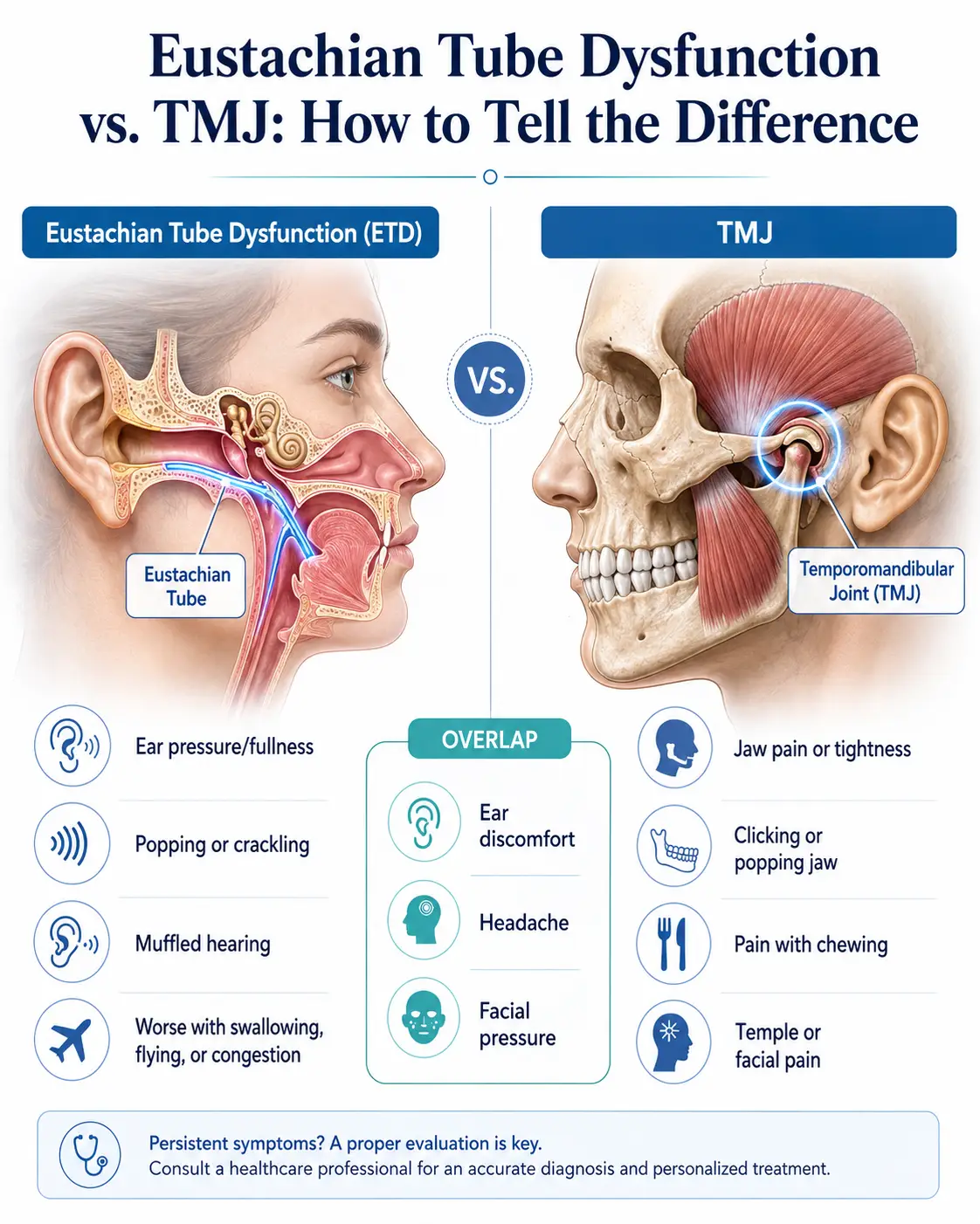
The jaw sits in the same neighborhood and on the same trigeminal nerve, and TMJ dysfunction produces ear fullness too. The sorting: jaw-driven ear symptoms travel with jaw clicking, pain on chewing, and tenderness in front of the ear; neck-driven symptoms travel with neck stiffness and skull-base pressure; and many people have both, because the upper neck, the jaw, and the ear all converge on the trigeminocervical complex. Our article on Eustachian tube dysfunction vs. TMJsorts the two in detail.
The Upper Cervical Connection
Everything above runs through the top of the neck, which is why our office focuses on the atlas and axis rather than the neck in general.
The atlas (C1) and axis (C2) sit at the junction of the skull and spine, directly beneath the brainstem where the trigeminocervical complex lives, and alongside the skull base where the tensor veli palatini and the cartilaginous Eustachian tube attach. The upper cervical nerve roots that feed the trigeminocervical complex exit right there. When the atlas is misaligned, three things follow: the C1–C3 nerve roots send distorted input into the shared relay continuously; the small muscles at the top of the neck stay under uneven tension, which is the stiffness and skull-base pressure patients feel; and the mechanics of the whole neck below are affected, producing the broader neck pain.
Correcting the position of the upper cervical spine addresses the input at its source — the same input that reaches the muscle opening your ear and the same input that refers sensation into the ear. Our neck pain guide covers the neck side in full, and our article on ear pressure and upper cervical chiropractic covers the ear side.
Upper Cervical Care at Lavender Family Chiropractic
When someone with ETD and neck pain comes to our Sarasota office, the first visit is an evaluation, not an adjustment.
History. The pattern of both symptoms — which came first, whether they move together, what provokes each, injury history, and what the ENT workup found.
Examination. Neurological screening; reproduction testing of the upper neck, which tells us quickly whether pressing on or moving the upper cervical spine reproduces the ear or head symptoms; range of motion; and palpation of the suboccipital muscles and the upper cervical joints.
Paraspinal infrared thermography. A non-invasive scan of the heat pattern along the spine, reflecting nervous-system activity — an objective baseline of irritation at the upper neck and a way to track change visit to visit.
3D cone-beam CT (CBCT) imaging of the upper cervical spine. The position of the atlas and axis relative to the skull, in three dimensions and to the degree — information a flat X-ray can’t give and an ENT workup doesn’t include.
Review of findings before any care.
Correction, if indicated. A precise, low-force procedure specific to your measurements — no twisting, no cracking, no rotation of the neck. Most patients describe it as pressure.
Remeasurement. Thermography after each visit and follow-up imaging when indicated.
Upper cervical care addresses the mechanical source the research points to — the neck — with measurement before and after, so progress is objective rather than guessed at. Results vary from person to person, and we tell you plainly what the findings show. Upper cervical care works alongside your ENT’s management; it addresses the input the ENT workup doesn’t examine.
If your ear and your neck have been bothering you together, call (941) 243-3729 or request an appointment online for an upper cervical evaluation.
What the Research Says
ETD defined and typed. Schilder and colleagues (2015) — the international consensus statement.
The tube is opened by the tensor veli palatini. Leuwer (2016) on the anatomy; Alper and colleagues (2012) measured the muscle’s activity against tube opening.
The trigeminal system and the upper neck share a brainstem relay. Bartsch and Goadsby (2003) on the trigeminocervical complex.
Ear symptoms are referred from the neck. Norris and Chen (2020) mapped secondary otalgia pathways including the cervical spine; Ramazani and colleagues (2023) on referred ear pain in primary care.
The upper neck’s position input affects head and sensory function. Kristjansson and Treleaven (2009).
Managing Both Day to Day
Posture at screens. Forward head posture loads the top of the neck and tightens the suboccipital muscles that sit beside the tube’s attachment. Screen at eye level, chin gently tucked, breaks every half hour.
Pillow. A neck held in a neutral line overnight. Side-sleepers need a fuller pillow; back-sleepers a lower one with neck support. No stomach sleeping.
Gentle swallowing and yawning, not forcing. Swallowing and yawning fire the tensor veli palatini naturally. Avoid forceful Valsalva (pinch-and-blow) more than occasionally; forced pressure can injure the eardrum.
Heat on the neck. A warm pack at the base of the skull eases the muscular component.
Allergy and sinus management with your physician. If inflammation is part of your ETD, treating it removes one input — and makes the neck component easier to see.
Don’t crack your own neck. Self-manipulation and rotational adjustments load the upper cervical joints you’re trying to quiet.
Serving Sarasota, Bradenton, Lakewood Ranch, and the Suncoast
Our office is on Whitfield Avenue in north Sarasota, minutes from the Manatee County line. We see patients with Eustachian tube dysfunction and neck pain from Sarasota, Bradenton, Lakewood Ranch, Palmetto, Parrish, Ellenton, Longboat Key, Siesta Key, Osprey, Nokomis, Venice, and North Port. If your ENT found the ear intact and your ear is still full, the top of the neck is the piece that hasn’t yet been examined.
Top 15 FAQs About Eustachian Tube Dysfunction and Neck Pain
1. Can neck problems cause Eustachian tube dysfunction? The muscle that opens the tube runs on trigeminal signal, and the trigeminal system shares its brainstem relay with the upper cervical nerves. An irritated upper neck disturbs that signal. The neck doesn’t block the tube; it affects the muscle that opens it.
2. Why does my ear feel full when my neck is tight? Two reasons: the disturbed signal to the tube’s opening muscle, and referred sensation — the ear receives sensory fibers from the upper cervical nerves, so neck irritation can be felt in the ear directly.
3. Can a pinched nerve in the neck cause ear pressure? Upper cervical nerve irritation can produce ear fullness and pressure through both pathways above. The radiology literature lists the cervical spine among the sources of referred ear symptoms.
4. How do I know if it’s my ear or my neck? If the ENT found the ear intact and the fullness travels with neck stiffness, skull-base pressure, or neck position, the neck is a factor. Reproduction testing at the first visit tells us quickly.
5. Why is it only one ear? Upper cervical misalignment is usually asymmetric, so the input to the trigeminocervical complex is uneven and the worse side gets the symptoms.
6. I had whiplash years ago. Could that be why? Yes. Whiplash disturbs the upper neck’s position and the input from it, and ear fullness is a recognized post-whiplash symptom that can surface long after the injury.
7. Does the Valsalva maneuver help? It can equalize a stuck ear temporarily. It doesn’t address a signal problem at the neck, and forcing it can injure the eardrum. Use it gently and occasionally.
8. Is it TMJ instead? It can be, or both. Jaw-driven ear fullness travels with jaw clicking and chewing pain; neck-driven with neck stiffness and skull-base pressure. Both converge on the same trigeminocervical relay.
9. Will balloon dilation fix it if the neck is the problem? Balloon dilation widens the tube — it addresses the tube itself. If the tube is intact and the signal to its muscle is the problem, widening it doesn’t address the source.
10. Can upper cervical care make my ear pop? The correction addresses the position of the upper neck, not the tube directly. When the neck is a factor, patients typically report the ear equalizing more normally over the following weeks as the input settles.
11. Is the first visit an adjustment? No. History, examination, thermography, and imaging if indicated, with findings reviewed before any care begins.
12. Should I see the ENT first? If you haven’t, yes — the ear itself needs to be examined, and that workup is what tells us the tube is intact. Bring the results.
13. How soon would I notice a difference? When the upper neck is a factor and it’s corrected, patients typically report changes in neck tightness first, then ear fullness, within the first several weeks. We measure before and after.
14. Can this cause headaches too? Yes — the ETD-headache connection runs through the same trigeminocervical wiring, and many patients have all three: ear fullness, neck pain, and headache.
15. Do you take insurance? Call the office and we’ll go through costs and coverage before anything begins.
Lavender Family Chiropractic in Sarasota, Florida offers a thorough upper cervical evaluation to find out whether we can help. Call (941) 243-3729 or request an appointment. 5899 Whitfield Avenue, Suite 107, Sarasota, FL 34243.
Related Articles
- Eustachian Tube Dysfunction: Our Complete Guide
- Neck Pain: Our Complete Guide
- Can Eustachian Tube Dysfunction Cause Headaches?
- Ear Fullness After a Car Accident
- Ear Pressure and Upper Cervical Chiropractic
- Eustachian Tube Dysfunction vs. TMJ
References
- Schilder AG, et al. Eustachian tube dysfunction: consensus statement on definition, types, clinical presentation and diagnosis. Clin Otolaryngol. 2015. PMID 26347263
- Leuwer R. Anatomy of the Eustachian Tube. Otolaryngol Clin North Am. 2016. PMID 27468634
- Alper CM, et al. Relationship between the electromyographic activity of the paratubal muscles and eustachian tube opening. Arch Otolaryngol Head Neck Surg. 2012. PMID 22801708
- Bartsch T, Goadsby PJ. The trigeminocervical complex and migraine: current concepts and synthesis. Curr Pain Headache Rep. 2003. PMID 12946290
- Norris CD, Chen RC. Secondary Otalgia: Referred Pain Pathways and Pathologies. AJNR Am J Neuroradiol. 2020. PMID 33093134
- Ramazani F, et al. Referred otalgia: Common causes and evidence-based strategies for assessment and management. Can Fam Physician. 2023. PMID 37963787
- Kristjansson E, Treleaven J. Sensorimotor function and dizziness in neck pain: implications for assessment and management. J Orthop Sports Phys Ther. 2009. PMID 19411769
Related Posts


Get our upper cervical articles first.
Patients Experience