Small-Fiber Neuropathy: Burning, Tingling, Normal EMGs — and the One Place No One Checked
By Dr. Rusty Lavender, D.C. — upper cervical chiropractor, Lavender Family Chiropractic, Sarasota, FL. Reviewed and updated October 2026.
You have burning feet, prickling hands, and electric jolts that come out of nowhere — and every test came back “normal.” Here is what almost no one tells patients from Sarasota to Lakewood Ranch to Bradenton: the burning is real, the nerves are misfiring, and the master control center for every sensory signal in your body sits at the top of your neck, in a region your entire workup probably never examined. Small-fiber neuropathy usually needs a proper medical cause found — and finding the cause is exactly why the upper cervical spine belongs on the list, because when the junction where your skull meets your spine is misaligned, it can irritate and dysregulate the very nervous system that decides how loud your nerves scream.
This article gives you the confident, cause-forward version. What small-fiber neuropathy actually is, why standard nerve testing is blind to it, how it is properly diagnosed, what drives it — and why the overlooked structural piece at the base of your skull deserves to be evaluated instead of ignored. Because you can chase the burning in your feet forever, but if the system that processes those signals is being irritated one inch below your skull, you are treating the smoke while the wiring problem goes unexamined.
This article is educational and not a substitute for medical advice. If you have a health condition, are pregnant, or take medications, talk with your physician before starting anything new.
What Small-Fiber Neuropathy Actually Is
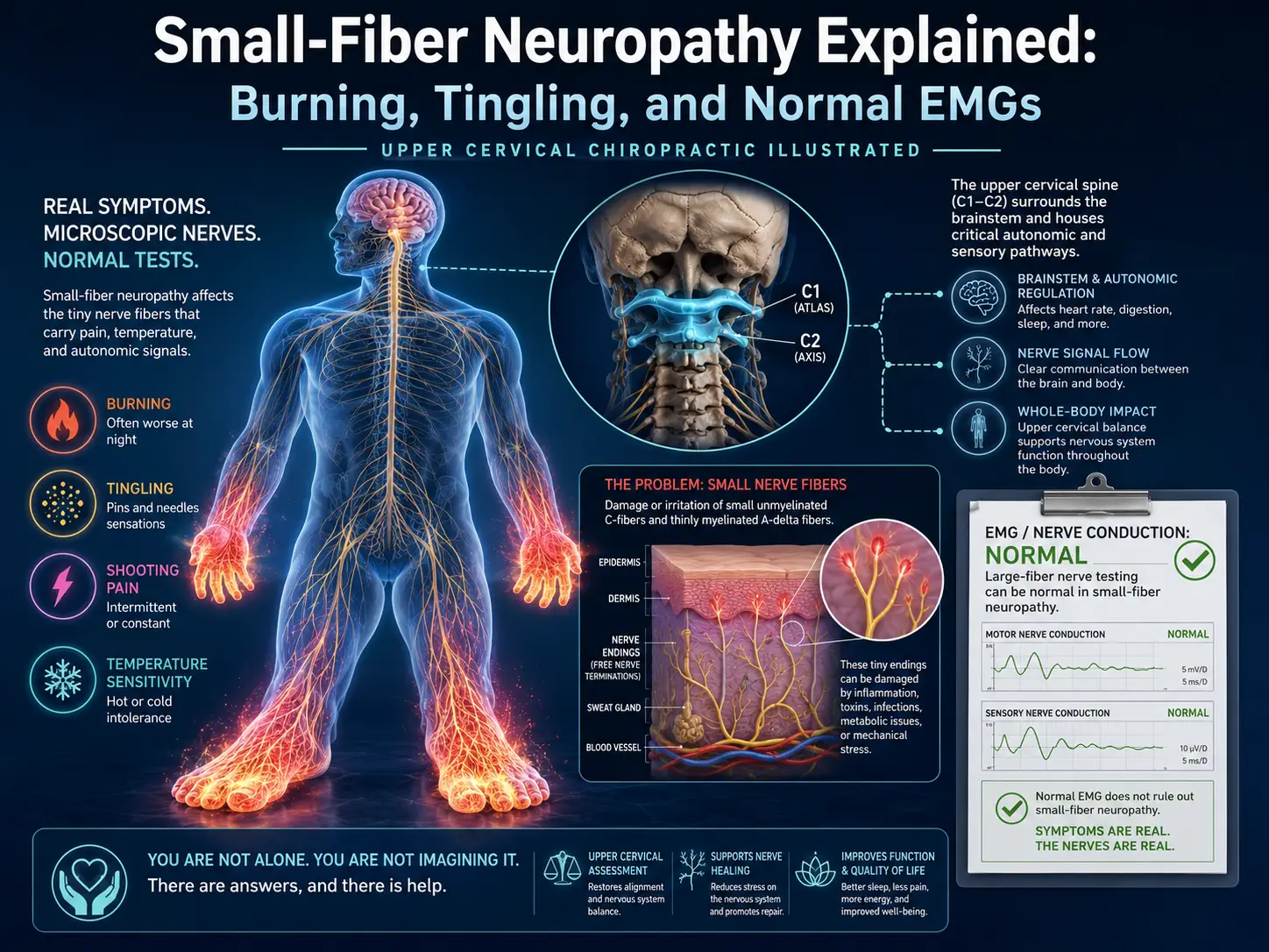
Your peripheral nervous system is built from nerve fibers of different sizes. The “large” fibers are thick, heavily insulated cables that carry signals for muscle strength, vibration sense, position sense (knowing where your foot is without looking), and the reflexes a doctor checks by tapping your knee. The “small” fibers are the thin ones — some lightly insulated, some bare — and they carry a completely different set of signals.
Small fibers do two main jobs:
- Sensory work. They carry pain, temperature (hot and cold), and certain kinds of touch. When they misfire, you feel burning, tingling, prickling, itching, or pain that seems wildly out of proportion to any injury.
- Autonomic work. Many small fibers are part of the autonomic nervous system — the automatic network that quietly regulates sweating, blood pressure, heart rate, digestion, and blood flow to the skin. When these fibers are stressed, people get abnormal sweating, lightheadedness on standing, dry eyes and mouth, or digestive changes.
Small-fiber neuropathy (SFN) is the condition in which these thin fibers become damaged, irritated, or degenerate. Because the small fibers are the ones affected, the classic symptoms are sensory and autonomic, while muscle strength usually stays intact — at least early on. That single fact explains most of the confusion patients live through, and we will keep coming back to it.
Here is the frame that changes everything: your peripheral nerves do not decide on their own how loud to fire. They report upward to the brainstem and upper spinal cord, the master relay that receives, processes, and regulates every sensory signal your body generates. How intensely you feel a burning foot is not set in the foot alone. It is modulated by a control center that sits, anatomically, at the very top of your neck.
What Small-Fiber Neuropathy Feels Like
No two people describe SFN in exactly the same words, but the patterns repeat. Putting language to what you feel is the first step toward being taken seriously.
Common Sensory Symptoms
- Burning pain, often in the feet — like the skin is on fire or you are walking on hot sand.
- Tingling and prickling, the “pins and needles” feeling, coming and going or staying constant.
- Electric-shock or stabbing sensations that shoot through the feet or hands without warning.
- Numbness, though people often describe a strange “wooden” or “thick” feeling rather than total loss of sensation.
- Allodynia, where things that should not hurt do hurt — bedsheets on the feet at night, socks, a light touch.
- Itching or crawling sensations on the skin with nothing visibly there.
The Classic Pattern
Small-fiber symptoms very often begin in the feet and toes — the parts of the body farthest from the spinal cord — and slowly climb upward over months or years. This is called a “length-dependent” pattern, because the longest nerves are affected first. In many people the hands become involved once the leg symptoms reach roughly knee level, producing the classic “stocking-and-glove” distribution.
Not everyone follows this map. Some people have patchy, non-length-dependent SFN, with symptoms in the face, trunk, arms, or scattered areas that ignore the usual bottom-up progression. This pattern is more often linked to autoimmune or inflammatory causes and is easy to dismiss precisely because it does not look “typical.”
Symptoms Are Often Worse at Night
A hallmark of small-fiber pain is that it intensifies in the evening and at night. The burning that hummed in the background during a busy day in Bradenton or Lakewood Ranch becomes loud and impossible to ignore the moment you lie down and the distractions fall away. Sleep disruption is one of the most wearing parts of living with SFN.
Autonomic Symptoms
Because small fibers also run the autonomic system, some people notice symptoms they would never have connected to a nerve problem:
- Lightheadedness or dizziness when standing up quickly
- Changes in sweating, either too much or too little
- Dry eyes and dry mouth
- Digestive changes such as bloating, early fullness, constipation, or diarrhea
- Changes in skin color or temperature in the hands and feet
- Bladder or sexual function changes
When autonomic features are prominent, that is a loud signal — it points toward specific underlying drivers and deserves careful evaluation. It is also a direct clue that the problem is one of regulation, and regulation is governed from the top of the neck.
Why Standard Nerve Tests Miss It Completely
Here is the part that blindsides patients. You go to a neurologist or your primary doctor, they order the standard nerve studies, the results come back normal, and you leave wondering whether anything is actually wrong.
The two standard tests are nerve conduction studies (NCS) and electromyography (EMG), together usually just called “the EMG.” They are genuinely valuable — but you have to understand what they measure.
Nerve conduction studies deliver a small electrical pulse to a nerve and measure how fast and how strongly the signal travels. The catch: this test primarily reads the large, well-insulated fibers, because those are the fast, strong signal carriers the equipment is built to detect. EMG measures the electrical activity of muscles, which reflects large motor nerves.
Do you see the problem? Standard NCS and EMG are essentially blind to the small fibers. In pure small-fiber neuropathy the large fibers are still healthy, so the large-fiber test reads normal — even while the small fibers are quietly degenerating and causing very real pain. A normal EMG does not rule out small-fiber neuropathy. It simply tells you the large fibers are, for the moment, working fine.
This is one of the most important messages in the whole field: a normal EMG is not proof that nothing is wrong. It is a normal result on a test that was never designed to see the fibers responsible for your symptoms. If your burning and tingling were dismissed on the basis of a normal EMG alone, that dismissal was incomplete — and it is worth a direct conversation with your physician about small-fiber-specific evaluation.
How Small-Fiber Neuropathy Is Properly Diagnosed
Because the standard tests miss it, real diagnosis takes a different approach. Good diagnosis rests on three legs: a careful clinical history and exam, specialized testing that actually looks at small-fiber function or structure, and a search for the underlying cause. Let’s take them one at a time.
1. Clinical History and Examination
A thoughtful clinician starts by listening. Where the symptoms started, how they spread, when they are worst, and what accompanies them carry enormous diagnostic information. On exam, a doctor may test your ability to feel pinprick and temperature, since those travel along small fibers. In classic SFN these small-fiber sensations are reduced while strength, reflexes, and vibration sense (large-fiber functions) stay normal. That mismatch is a strong clue.
2. Skin Biopsy and Intraepidermal Nerve Fiber Density (IENFD)
The test most often described as the reference standard for confirming SFN is a skin biopsy used to measure intraepidermal nerve fiber density, or IENFD. It sounds more intimidating than it is. A clinician uses a small punch tool to take a tiny sample of skin, usually about three millimeters across, typically from the lower leg above the ankle. A specialized lab stains and counts the thin nerve fibers within the skin. In SFN, the number of these tiny nerve endings is reduced compared with established normal values matched for age and sex.
The beauty of this test is that it looks directly at the small fibers — the very fibers standard EMG cannot see. It is minimally invasive, generally well tolerated, and usually needs no stitches. If your workup has not included it and SFN is suspected, it is a reasonable thing to ask your neurologist about.
3. Quantitative Sensory Testing (QST)
Quantitative sensory testing measures how well you perceive specific sensations — warmth, cold, and heat-pain — carried by small fibers. A device applies carefully controlled stimuli and you indicate when you feel them. Because it depends on your responses, QST is “psychophysical”: it reflects the whole pathway from nerve to brain to your report. It is useful alongside, rather than instead of, skin biopsy.
4. Autonomic Testing
When autonomic symptoms are present, additional tests can assess the small autonomic fibers — sweat testing (such as QSART), tilt-table testing for blood pressure and heart rate responses to position change, and other cardiovascular reflex tests. These are especially useful when lightheadedness, sweating changes, or digestive symptoms are part of the story.
5. The Search for a Cause
Diagnosing SFN is only half the job. The other half — the more important half — is finding out why. This is where the workup either succeeds or quietly stops short. And this is exactly where the upper cervical spine gets overlooked.
Common Drivers of Small-Fiber Neuropathy
Most SFN traces back to a systemic driver, and treating the burning feet without finding it is like mopping the floor while the faucet runs. Here are the categories that come up most.
Diabetes and Prediabetes (Impaired Glucose Metabolism)
Disordered blood sugar is among the most common drivers, and this includes not only full diabetes but prediabetes and impaired glucose tolerance. Small-fiber damage can be one of the earliest signs of a blood-sugar problem, sometimes appearing before diabetes is ever diagnosed. That is why fasting glucose, hemoglobin A1c, and sometimes an oral glucose tolerance test are cornerstones of the SFN workup.
Vitamin and Nutritional Factors
Vitamin B12 deficiency is a classic, checkable one, which is why B12 is standard bloodwork. Other B vitamins — and excessive intake of vitamin B6 — can also play a role. Simple to test, often simple to address, and never worth overlooking.
Autoimmune and Inflammatory Conditions
The immune system can attack small nerve fibers, either directly or as part of a broader condition — Sjögren’s syndrome(classically dry eyes and mouth), lupus, sarcoidosis, celiac disease, and others. Some people have immune-driven SFN without a formally named disease. Autoimmune causes are especially worth pursuing when the pattern is patchy, non-length-dependent, or autonomic-heavy.
Thyroid, Kidney, and Other Metabolic Conditions
Thyroid dysfunction, chronic kidney disease, and other metabolic disturbances can all contribute and are typically screened during a complete evaluation.
Infections, Medications, and Genetic Causes
Certain infections (including HIV and hepatitis C) and some medications (particularly certain chemotherapy agents) can damage small nerves. In a subset of people — especially early-onset or unexplained cases — inherited conditions such as sodium-channel gene variants or Fabry disease are at play.
“Idiopathic” Too Often Means “No One Examined the Neck”
Despite a thorough search, a meaningful proportion of SFN gets labeled idiopathic — no clear cause found. Here is the honest, cause-forward truth patients rarely hear: “idiopathic” is a statement about how far the search went, not proof that no driver exists. And in an overwhelming number of workups, one region is never examined at all — the upper cervical spine, the junction where your skull balances on the atlas (C1) and axis (C2), inches from the brainstem and upper spinal cord that process and regulate every sensory signal your body produces. If the master relay for your sensory nervous system was never looked at, the search was not finished. “Idiopathic” so often means “no one checked the one place that could be driving how these signals are regulated.”
The Overlooked Driver: How the Upper Neck Can Dysregulate Your Nervous System
Here is where nearly every article on this topic stops short, and it is the most important part. The workup above assumes the burning in your feet is a purely local, downstream problem. But sensory intensity is not set only in the foot — it is processed and modulated upstream, in the brainstem and upper spinal cord.
Consider the anatomy. The atlas (C1) and axis (C2) are the two most mobile vertebrae in the spine, carrying the entire weight of your head and held mostly by ligaments rather than interlocking bone. They surround the brainstem and the top of the spinal cord — the master relay for all sensory and autonomic signaling. When that top segment shifts out of alignment, the surrounding tissues, muscles, and structures at the skull base come under abnormal tension and stress, at the exact junction where sensory and autonomic traffic is routed and regulated. The plausible consequence is not a simple neck ache. It is ongoing irritation of the very system that decides how your nerve signals are processed, gated, and amplified — which can keep an already-sensitized system locked in overdrive no matter what is happening down in your feet.
There is an autonomic and blood-flow angle too. Small fibers help regulate blood flow to the skin, and the autonomic control that governs vascular tone runs through this same upper cervical region. When the autonomic side of the nervous system is chronically dysregulated, the small fibers that depend on healthy blood flow and stable autonomic input are working in a hostile environment. That is a mechanism worth taking seriously — and evaluating — rather than waving away.
This also explains a pattern we see constantly: people whose nerve symptoms trace back to a head or neck injury. A concussion, a whiplash, a car accident, or years of forward-head posture hunched over a screen loads and destabilizes the very region the brainstem and upper cord occupy. If your burning, tingling, or autonomic symptoms started or worsened after something happened to your head or neck, that is a loud clue that a structural driver may be sitting one inch below your skull — in a region your workup almost certainly never examined. None of this replaces finding a metabolic cause. It means the upper cervical spine belongs on the list of things to check, not left off it.
How We Evaluate the Structural Piece at Lavender Family Chiropractic
At Lavender Family Chiropractic in Sarasota, we focus exclusively on the upper cervical spine — the atlas, the axis, and the junction where the skull meets the neck, the same region that houses the master relay for your sensory and autonomic nervous system. Our approach is built on measurement, not guesswork. We use 3D CBCT imaging to see your upper cervical alignment in three dimensions, and paraspinal infrared thermography to read how your nervous system is actually behaving along the spine — directly relevant when the whole question is nerve signaling and autonomic regulation. When a correction is warranted, we use the Knee Chest Upper Cervical technique: precise, gentle, and low-force, with no twisting, cracking, or popping. For an already-sensitized nervous system, that gentleness is the entire point.
The goal is simple: give an irritated, overloaded nervous system a calmer structural environment. Keep your physician, keep the metabolic workup, keep chasing the blood sugar and B12 and thyroid answers — those matter enormously. What we add is attention to the one region that sits at the crossroads of your spine and the sensory control center, the piece the standard neuropathy workup leaves out. If you have burning feet, normal EMGs, and a stack of “idiopathic” labels, that is the evaluation you have been missing.
Serving Sarasota, Lakewood Ranch & Bradenton
Patients travel to our Sarasota office from across the region — Sarasota, Lakewood Ranch, Bradenton, University Park, Palmetto, and the greater Sarasota-Manatee area — because so few clinics anywhere look at the upper cervical spine when nerve symptoms will not settle. If you live in Sarasota, Lakewood Ranch, or Bradenton and you have been told your nerve tests are “normal” while your feet burn every night, you do not have to accept a dead end. We are right at the corner of University and Whitfield, minutes from Lakewood Ranch and a short drive from downtown Bradenton, and we would be glad to evaluate whether your upper neck is part of your picture.
→ Call Lavender Family Chiropractic at (941) 243-3729 to schedule a consultation. We are at 5899 Whitfield Avenue, Suite 107, Sarasota, FL 34243 — serving Sarasota, Lakewood Ranch, and Bradenton.
Red Flags: When to Seek Prompt Medical Care
Most small-fiber neuropathy develops slowly, but certain symptoms warrant a timely call to your physician or, when severe, urgent medical attention. Please do not wait to have these evaluated:
- Rapidly progressive weakness, or any new muscle weakness, especially if it is spreading quickly. Pure SFN typically spares strength, so new weakness suggests something else that needs prompt assessment.
- Prominent or worsening autonomic symptoms, such as fainting, severe lightheadedness on standing, or a racing or irregular heartbeat.
- Sudden, severe, or rapidly spreading symptoms, rather than the slow creep that is more typical.
- New bladder or bowel changes accompanying the nerve symptoms.
- Any signs of infection, foot wounds, or ulcers, particularly if you have reduced sensation and may not feel an injury — and, if you have diabetes, any signs of a diabetes complication warrant urgent care.
When in doubt, reach out to your medical provider. Timely evaluation of these features can make a real difference.
What the Research Says
I want to ground this in the published literature rather than opinion, so here is what the peer-reviewed evidence tells us about diagnosing and understanding small-fiber neuropathy.
The reference-standard role of skin biopsy is well established. A joint guideline from the European Federation of Neurological Societies and the Peripheral Nerve Society concluded that measuring intraepidermal nerve fiber density through skin biopsy is a reliable and validated method for confirming small-fiber neuropathy, and it provided practical recommendations for how the biopsy should be performed and interpreted (EFNS/PNS guideline on skin biopsy, European Journal of Neurology, 2010).
The framework for how we define and diagnose SFN in the clinic was significantly shaped by research proposing formal diagnostic criteria that combine clinical signs and symptoms with objective testing. An influential 2008 study helped establish how symptoms, examination findings, and neuropathological confirmation via skin biopsy fit together into a coherent diagnostic approach (Devigili and colleagues, Brain, 2008). More than a decade later, that same line of research was revisited and refined for both clinical practice and research settings, sharpening the criteria so clinicians can diagnose SFN more consistently (Devigili and colleagues, Brain, 2019).
For clinicians on the front lines, a practical review offered an office-based approach to recognizing and evaluating small-fiber neuropathy, emphasizing the value of a careful history, the appropriate use of specialized testing, and a systematic search for underlying and often treatable causes (Cleveland Clinic Journal of Medicine, 2018). And a broad review in a leading neurology journal captured the honest reality that diagnosing small-fiber neuropathy remains genuinely challenging, that no single test is perfect, and that combining clinical judgment with the right investigations gives the most accurate picture while an underlying cause is pursued (Lancet Neurology, 2017).
Taken together, this body of work supports the core messages here: standard EMG does not evaluate small fibers, skin biopsy with IENFD is the most direct way to confirm the diagnosis, and finding the underlying cause is central to real care — which is exactly why a region that governs how sensory signals are processed should not be left unexamined.
Top Questions
If my EMG was normal, does that mean I don’t have neuropathy? Not at all. Standard EMG and nerve conduction studies measure the large nerve fibers. In small-fiber neuropathy the large fibers are typically normal, so those tests come back normal even though the small fibers are affected. A normal EMG does not rule out SFN. If your symptoms fit the small-fiber picture, ask your physician about small-fiber-specific testing such as skin biopsy — and about examining the upper neck.
What is the most direct way to confirm small-fiber neuropathy? A skin biopsy that measures intraepidermal nerve fiber density (IENFD) is widely regarded as the reference-standard test. It uses a tiny skin sample, usually from the lower leg, to directly count the small nerve endings in the skin. Quantitative sensory testing and autonomic testing add supporting information.
Why would my neck matter for burning in my feet? Because your feet do not decide on their own how loudly to fire. Sensory signals report to the brainstem and upper spinal cord — the master relay that processes and regulates them — and that relay sits at the top of your neck, surrounded by the atlas and axis. When that junction is misaligned, it can irritate and dysregulate the very system that governs how your nerve signals are amplified. That is why the upper cervical spine is worth evaluating, especially when the cause is otherwise called “idiopathic.”
My symptoms started after a car accident or concussion. Does that matter? It matters a great deal. Trauma to the head and neck loads and destabilizes the exact region the brainstem and upper cord occupy. If your nerve symptoms began or worsened after a neck injury, the upper cervical spine is a driver worth evaluating rather than assuming away.
What bloodwork should I ask about? Common tests in an SFN evaluation include fasting glucose and hemoglobin A1c (and sometimes an oral glucose tolerance test), vitamin B12, thyroid function, and screening for autoimmune conditions such as Sjögren’s syndrome. Your physician will tailor the workup to your history.
Why are my symptoms worse at night? Many people with SFN notice burning and tingling intensify in the evening and at night, when daytime distractions fade and you lie still. This is a common, recognized pattern. Good sleep habits and, when appropriate, physician-guided symptom management help.
Is the adjustment forceful? No — precise, gentle, low-force, with no twisting, cracking, or popping.
Is any of this a substitute for medical care? No. This article is educational. If you have symptoms, a diagnosed condition, are pregnant, or take medication, work with your physician. Upper cervical care complements good medical care — it does not replace it.
Find the Cause — and Check the One Place No One Looked
Your symptoms are real, there are specific tests designed to find small-fiber neuropathy, and identifying the underlying driver is the path to meaningful help. But if you have done the bloodwork, sat through the “normal” EMG, and collected the word “idiopathic,” ask the question the standard workup never does: has anyone examined the one region that governs how your nervous system processes these signals?
For a great many people from Sarasota, Lakewood Ranch, and Bradenton, that upper cervical evaluation is exactly what has been missing — and it is the one place no one checked.
Let us look at what the standard workup skips. Call (941) 243-3729 or book a consultation. Find the cause — and make sure the master relay is free to do its job.
Lavender Family Chiropractic (NeckWise North Sarasota) · 5899 Whitfield Avenue, Suite 107, Sarasota, FL 34243 · (941) 243-3729 · Serving Sarasota, Lakewood Ranch & Bradenton
This article is for general educational purposes only and is not medical advice, diagnosis, or treatment. Small-fiber neuropathy should be evaluated by a qualified physician. Please consult your physician about your individual symptoms, testing, and care, and seek prompt medical attention for red-flag symptoms such as rapidly progressive weakness or significant autonomic changes.
Related Articles
Related Posts


Get our upper cervical articles first.